
Dermatology Procedures · Patient Guide
Types of Skin Biopsy
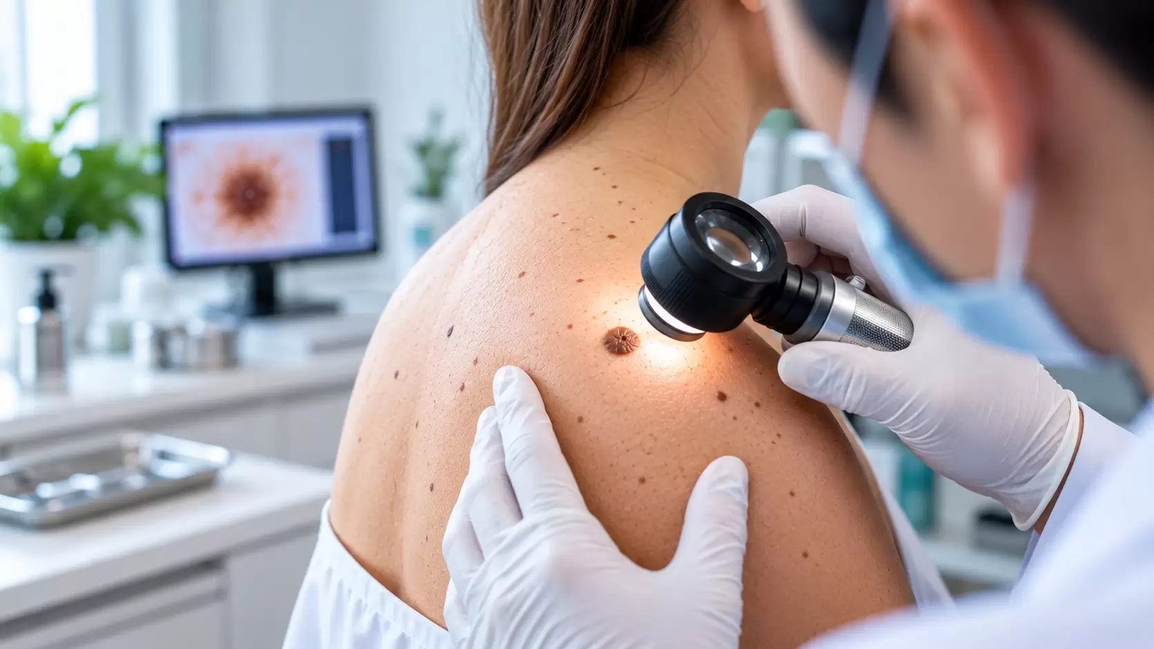
Punch, shave and excisional biopsies collect skin tissue in different ways. The appropriate method depends on the lesion, its depth, its location and the diagnostic question.
A shave biopsy samples mainly superficial skin, a punch biopsy removes a small cylindrical full-thickness sample, and an excisional biopsy removes an entire lesion when appropriate. Your dermatologist selects the technique that is most likely to provide the pathology information needed.
Understanding the procedure
Why are there different biopsy types?
A skin biopsy is not one single procedure. It is a group of techniques used to obtain tissue for microscopic examination. A dermatologist may recommend biopsy when the appearance of a rash, growth, mole or other skin change does not provide enough information on its own, or when microscopic examination is important for confirming the diagnosis.
The American Academy of Dermatology explains that biopsy technique varies according to the suspected condition and the size or characteristics of the growth. DermNet similarly notes that choosing the correct biopsy type and site is important because the sample must contain the tissue features the pathologist needs to evaluate.
This is why one patient may need a superficial shave while another needs a deeper punch or removal of the entire lesion. The method is selected for a clinical reason rather than simply because one technique is universally better.
The three common approaches
Punch vs shave vs excisional biopsy
Punch biopsy
A circular punch instrument removes a small cylindrical core containing the epidermis, dermis and usually deeper tissue. It is often useful when the pathologist needs to assess the architecture across several skin layers.
Shave biopsy
A blade removes a thin or tangential portion of a superficial lesion. Because the sample is shallower, it is generally selected when the relevant diagnostic features are expected near the skin surface.
Excisional biopsy
The entire visible lesion is surgically removed, usually with a scalpel. The wound commonly requires stitches. Excision can provide the pathologist with the whole lesion for examination.
Punch biopsy
When is a punch biopsy useful?
A punch biopsy uses a small, tube-shaped cutting instrument. After the area is cleaned and numbed with local anaesthetic, the dermatologist rotates the punch through the skin to obtain a cylindrical sample. According to DermNet, commonly used punches are only a few millimetres across, yet they can provide a full-thickness specimen containing multiple skin layers.
This depth makes punch biopsy useful for many inflammatory skin diseases and for selected lesions where information below the surface is important. The biopsy site matters as much as the instrument: depending on the suspected condition, the dermatologist may choose the centre, edge or nearby skin to obtain the most informative tissue.
A small punch site may sometimes heal without sutures, while another may require one or more stitches. The decision depends on the punch size, body location and wound characteristics. Learn more about the overall process on our Skin Biopsy in Chennai treatment page.
What does a punch biopsy show?
Because a punch can include the epidermis, dermis and sometimes subcutaneous tissue, it allows the pathologist to evaluate changes occurring at different depths. This can be particularly useful when the pattern and distribution of inflammation within the skin are diagnostically important.
Shave biopsy
What is a shave biopsy?
In a shave biopsy, a dermatologist uses a blade or specialised shave instrument to remove a superficial portion of the skin. The technique is often considered for raised or superficial lesions when a deep full-thickness specimen is not required.
DermNet notes that shave biopsy generally does not include the full thickness of the skin. That is useful in appropriate cases but can also be a limitation when determining the depth of a process is essential. The dermatologist therefore considers the suspected diagnosis before deciding whether a shave sample is suitable.
Shave biopsy wounds are often left to heal without stitches. Wound care remains important: follow the instructions provided for cleansing, protection and dressing of the biopsy site.
The two techniques sample tissue differently. A shave emphasizes surface area and superficial tissue, whereas a punch provides a narrower but deeper cylindrical sample.
Excision
What is an excisional biopsy?
An excisional biopsy removes the entire visible lesion rather than taking only a portion. A scalpel is typically used, and the resulting wound is generally closed with sutures. This approach may be considered when examination of the complete lesion is important and when its size and location make complete removal appropriate.
An incisional biopsy is related but different: instead of removing the entire lesion, it removes only a representative section. The distinction is important because the objective may be diagnosis alone or diagnosis together with complete removal of a small lesion.
Not every mole or growth should automatically be removed by the same technique. If you are concerned about a mole, see our information on mole assessment and removal in Chennai. A dermatologist can first assess the lesion and discuss whether biopsy is indicated and which method is appropriate.
How the choice is made
Which type of biopsy might you need?
The choice is based on what the dermatologist is trying to learn from the sample. Factors can include the suspected diagnosis, whether the condition is superficial or deep, the size and shape of the lesion, its body location, whether the whole lesion needs examination, and the type of tissue the pathologist needs.
For inflammatory rashes, selecting the right part and age of a lesion can affect how informative the pathology is. For growths or suspected tumours, depth and the relationship between abnormal and surrounding tissue may be important. In some situations, the dermatologist may need more than one specimen or a specimen specifically prepared for a specialised laboratory test.
This clinical selection is one reason a biopsy should be viewed as part of a diagnostic pathway rather than as an isolated procedure. If you are unsure why biopsy has been recommended, ask what question the biopsy is intended to answer and what the result may change about your treatment.
Does the biopsy type affect the scar?
Any procedure that breaks the skin can leave a mark or scar. Its appearance varies with the biopsy method, wound size, body site, individual healing tendency and aftercare. A punch or excision may require sutures, while a shave commonly heals without them. Your dermatologist can explain the expected wound and aftercare before the procedure.
Does a biopsy always mean cancer is suspected?
No. Skin biopsy is used to diagnose many conditions, including inflammatory diseases, infections, blistering disorders and skin cancers. The American Academy of Dermatology specifically notes that dermatologists use biopsy for many different skin conditions, not only cancer. For a detailed clinical overview of techniques, see DermNet’s skin biopsy guide.
After the sample is taken
What happens after a biopsy?
The removed tissue is sent for histopathology, where it is processed and examined under a microscope. The pathology findings are interpreted alongside the clinical information supplied by the doctor. Depending on the result, the biopsy may confirm the suspected diagnosis, suggest another diagnosis or indicate that additional assessment is needed.
The biopsy wound also needs time to heal. Keep the site clean and follow the dressing instructions provided by your doctor. Contact the clinic if you have concerns such as increasing pain, spreading redness, significant swelling, persistent bleeding or discharge.
If you want a broader overview before undergoing the procedure, read What Is a Skin Biopsy and Why Is It Done?. Our next patient guide will cover how to prepare for a skin biopsy.
Frequently asked questions
Skin biopsy FAQs
What are the main types of skin biopsy?
Common methods include punch, shave, incisional and excisional biopsy. Other sampling techniques may also be used depending on the lesion and diagnostic purpose.
Is punch biopsy deeper than shave biopsy?
Generally, yes. A punch obtains a cylindrical full-thickness sample through multiple skin layers, whereas a standard shave biopsy mainly samples superficial tissue.
Does a punch biopsy need stitches?
Some punch biopsy wounds require a suture while very small wounds may heal without one. The decision depends on the size and location of the biopsy and other clinical factors.
Does an excisional biopsy remove the whole lesion?
By definition, an excisional biopsy removes the entire lesion being sampled. An incisional biopsy removes only part of a larger lesion.
Who decides which skin biopsy technique is appropriate?
The dermatologist selects the biopsy method after examining the lesion and considering the suspected diagnosis, required tissue depth, body site and other clinical factors.
Visit us in OMR
Let’s talk about
your skin concerns.
Book a consultation at Ram Skin Clinic in Thoraipakkam, Chennai.
#48, Second Floor, Flat E, Best Towers,Okkiampet, Thoraipakkam, OMR,
Chennai – 600097. Above Domino’s Pizza.