
Understanding Marks Present From Birth
Common Types of Birthmarks: Vascular & Pigmented Birthmarks Explained
Birthmarks are areas of skin that look different from the surrounding skin and are present at birth or appear shortly afterwards. They vary widely in colour, shape, size and behaviour, and different types need different follow-up.
What exactly is a birthmark?
A birthmark is a visible skin marking that develops before birth or shortly afterwards. It may be flat or raised and can be pink, red, purple, blue-grey, tan, brown or darker than the surrounding skin.
Some birthmarks fade during childhood, while others remain throughout life. Their behaviour depends on the specific type rather than simply their colour.
Two broad groups of birthmarks
Vascular birthmarks
These involve blood vessels. Examples include infantile haemangiomas, salmon patches and capillary malformations such as port-wine stains.
Pigmented birthmarks
These involve pigment-producing cells or pigment within the skin. Examples include congenital melanocytic naevi, café-au-lait spots and dermal melanocytosis.
There are also other developmental naevi derived from epidermal, sebaceous, connective-tissue and other skin structures.
1. Infantile haemangioma
Infantile haemangiomas are vascular growths that often appear at birth or within the first weeks of life. A superficial haemangioma can look bright red and raised, while a deeper lesion may appear bluish or skin-coloured.
They commonly grow rapidly during early infancy and then gradually shrink over several years. Many can be observed, but haemangiomas near the eye, nose, mouth or other functionally important areas may need early treatment.
2. Salmon patch
Salmon patches are common flat pink-red vascular marks. They are often seen on the forehead, eyelids, between the eyebrows or at the back of the neck.
Facial salmon patches commonly become less noticeable with time. They may look more prominent when a baby cries, becomes warm or strains.
3. Port-wine stain
A port-wine stain is a capillary malformation that appears as a flat pink, red or purple patch. Unlike an infantile haemangioma, it does not naturally go through a growth-and-involution cycle.
Port-wine stains generally persist and may darken or thicken with age. Facial lesions in certain distributions can sometimes be associated with other conditions and warrant assessment.
4. Café-au-lait spots
Café-au-lait spots are flat, evenly pigmented tan-to-brown patches with clearly defined borders. A single spot is common and is usually harmless.
Multiple café-au-lait spots can sometimes be associated with genetic conditions. The AAD recommends dermatology assessment when a child has six or more or develops freckle-like spots around them.
5. Congenital melanocytic naevus
A congenital melanocytic naevus is a mole present at birth or developing shortly afterwards. It can range from a small brown spot to a much larger lesion and may be smooth, raised, hairy or have a more textured surface.
Most small congenital moles remain benign. Larger congenital naevi require more individualised follow-up because melanoma risk increases with size and other features.
For a related comparison, see Birthmark vs Mole once available in this series.
6. Dermal melanocytosis
Dermal melanocytosis, historically called a Mongolian spot, appears as a blue-grey patch that can resemble a bruise. It most commonly occurs over the lower back or buttocks but can occur elsewhere.
These marks are particularly common in Asian and other darker skin tones and often fade during early childhood.
7. Naevus sebaceous
A naevus sebaceous is a developmental birthmark that most often occurs on the scalp or face. In infancy it may appear as a slightly raised, hairless yellowish patch.
It can become thicker or more textured around puberty and generally does not disappear spontaneously. Dermatology review helps establish the diagnosis and follow-up plan.
8. Hypopigmented birthmarks
Some birthmarks are lighter rather than darker than the surrounding skin. A hypopigmented macule or naevus depigmentosus may appear as a pale patch from birth or early life.
Multiple or unusual light patches can occasionally be associated with other conditions, so the overall pattern matters more than colour alone.
Do birthmarks change as a child grows?
Yes. Some birthmarks grow proportionally with the child, some such as infantile haemangiomas can enlarge rapidly before shrinking, and others may darken or thicken over time.
Knowing the expected behaviour of the specific birthmark makes it easier to distinguish normal evolution from a change that needs reassessment.
Which birthmarks tend to fade?
Many salmon patches fade, and most infantile haemangiomas eventually involute substantially. Dermal melanocytosis also commonly becomes less visible during childhood.
Port-wine stains, café-au-lait spots and most congenital melanocytic naevi generally persist, although their appearance may change as the child grows.
Which birthmarks should be checked?
Rapid growth or unexpected change
A lesion growing unusually quickly or changing colour, border or texture should be reassessed.
Pain, ulceration or bleeding
A birthmark that breaks down, becomes painful or bleeds may need wound care or treatment.
Important location
Marks near the eye, nose, mouth or other structures may affect function depending on their type and size.
Multiple lesions
Numerous café-au-lait spots or other specific patterns can occasionally point to an associated condition.
How does a dermatologist identify the type?
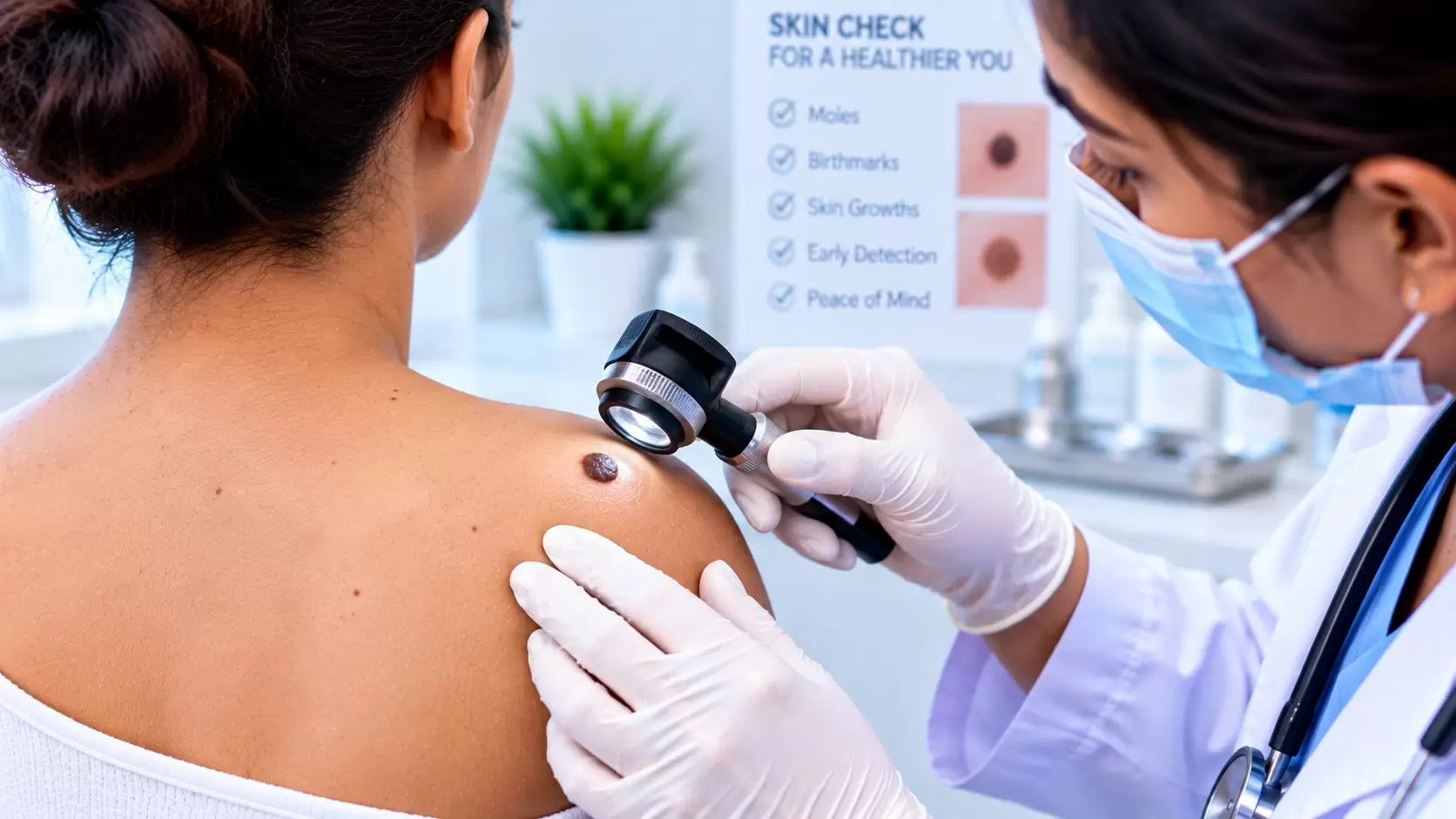
Many birthmarks can be diagnosed from their appearance, location and history. The dermatologist may use dermoscopy or a Wood’s lamp for closer examination.
Imaging, biopsy or other investigations are reserved for selected cases when the diagnosis is uncertain or when the birthmark’s type or distribution suggests deeper involvement or an associated condition.
Do all birthmarks need treatment?
No. Most birthmarks are harmless and can simply be monitored. Treatment is considered when a lesion may interfere with function, has complications, carries a specific medical risk or causes significant cosmetic or psychosocial concern.
Treatment also depends on the type. For example, propranolol may be used for selected infantile haemangiomas, while pulsed-dye laser can be considered for port-wine stains.
When should you see a dermatologist?
Birthmarks should be assessed when the diagnosis is uncertain, the mark grows rapidly or changes unexpectedly, it ulcerates or bleeds, or it is positioned where it could affect vision, feeding, breathing or another function.
Assessment is also appropriate for large congenital moles, multiple café-au-lait spots or when parents have concerns about how a mark is evolving.
For evaluation, visit Birthmark Assessment in Chennai.
Frequently asked questions
What are the main types of birthmarks?
Birthmarks are commonly grouped into vascular birthmarks involving blood vessels and pigmented birthmarks involving pigment-producing cells. Other developmental skin naevi can also be present at or shortly after birth.
Do all birthmarks disappear with age?
No. Some infantile haemangiomas and salmon patches often fade, while port-wine stains, café-au-lait spots and most congenital moles generally persist.
Is a congenital mole a birthmark?
Yes. A congenital melanocytic naevus is a mole present at birth or appearing shortly afterwards and is a type of pigmented birthmark.
Are birthmarks dangerous?
Most birthmarks are harmless, but some need assessment because of their type, size, location, number or changes over time.
When should a birthmark be checked by a dermatologist?
Assessment is appropriate when a birthmark grows rapidly, changes unexpectedly, becomes painful, ulcerates or bleeds, affects an important structure such as the eye or mouth, or when its diagnosis is uncertain.
Visit us in OMR
Not sure what type of birthmark it is? Start with an accurate assessment.
Book a dermatologist consultation at Ram Skin Clinic in Thoraipakkam, Chennai.
#48, Second Floor, Flat E, Best Towers,Okkiampet, Thoraipakkam, OMR,
Chennai – 600097. Above Domino’s Pizza.