
Mole Assessment & Removal
Mole Removal Methods
Moles may be removed because they are suspicious, repeatedly irritated or unwanted for cosmetic reasons. The appropriate method depends on how the mole looks, where it is located and whether tissue needs to be examined under a microscope.
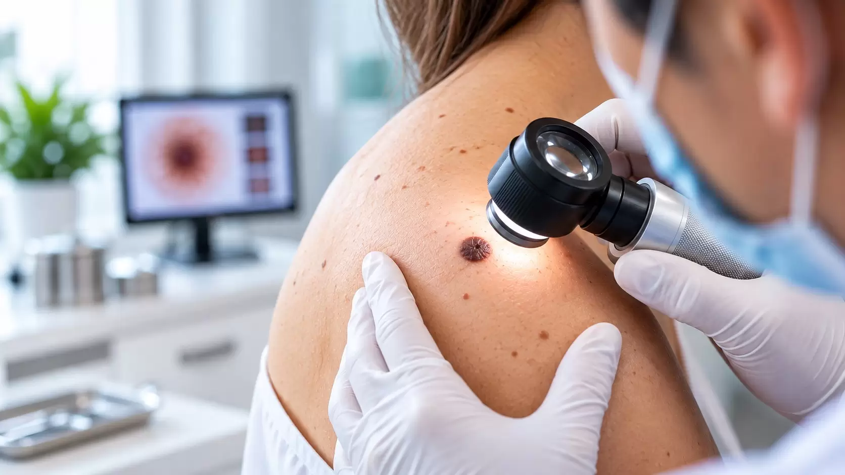
Most moles are benign and do not need to be removed. A dermatologist may recommend removal when a mole has concerning features, catches on clothing or a razor, becomes repeatedly irritated, or when a patient wants a benign mole removed for cosmetic reasons. Before choosing a technique, the first step is deciding whether the lesion is clinically typical or needs diagnostic assessment.
If you have a new or changing lesion, read when to get a mole checked and our guide to a normal mole vs suspicious mole. For in-person assessment, see our Mole Assessment & Removal in Chennai service page.
Why the diagnosis comes first
A removal technique should not be chosen purely on appearance or convenience. A suspicious pigmented lesion may need to be removed in a way that allows a pathologist to examine the architecture and depth of the lesion. Histopathology is the definitive method used to diagnose melanoma.
Common mole removal methods
Surgical excision
The dermatologist removes the mole with a scalpel, usually including the full depth required for the planned procedure. The wound is then closed with stitches when necessary. Excision is particularly important when complete tissue assessment is required. The specimen can be sent for histopathology.
Surgical shave removal
For selected raised moles that appear benign, a dermatologist may use a surgical blade to remove the protruding portion of the lesion. Depending on the depth, the area may heal without stitches. Shave removal is not interchangeable with excision for every pigmented lesion.
The American Academy of Dermatology describes both surgical excision and surgical shave as commonly used dermatologist-performed mole-removal techniques. DermNet similarly notes that flat or suspicious melanocytic naevi may require excision biopsy, while selected protruding benign-appearing naevi may be suitable for shave biopsy.
What about laser or cautery?
Some benign skin lesions can be treated with destructive procedures, and selected melanocytic naevi may sometimes be treated for cosmetic reasons after specialist assessment. However, destroying tissue means there may be little or no specimen available for microscopic examination. That makes diagnosis the priority whenever there is uncertainty about a pigmented lesion.
For this reason, a dermatologist should first establish what the lesion is. A treatment that is appropriate for an obviously benign raised mole may be inappropriate for a lesion with asymmetry, irregular borders, colour variation, recent evolution or other suspicious features.
Will mole removal leave a scar?
Any procedure that cuts or removes skin can leave a scar. The final appearance depends on the method, mole size and depth, body site, individual healing tendency and aftercare. Excision creates a linear surgical wound when stitches are used, while shave removal usually creates a more superficial wound that heals from the base upward.
A dermatologist balances complete or diagnostically adequate removal against the expected scar. Cosmetic outcome is important, but it should not override the need for proper assessment when cancer is a possibility. Our next article in this cluster covers mole removal and scarring in more detail.
What happens after removal?
The area is usually covered with a dressing and you will receive wound-care instructions based on the procedure. If stitches are used, your dermatologist will explain when they should be reviewed or removed. A specimen may be sent to a pathology laboratory, particularly when diagnosis is part of the reason for removal.
Do not assume that a mole has been permanently dealt with simply because the visible portion is gone. If pigmentation or a growth returns at a removal site, have it reassessed rather than trying to remove it again yourself.
For a broader explanation of diagnostic sampling, see our Skin Biopsy in Chennai page and What Is a Skin Biopsy?.
Choosing the right method
There is no single mole-removal method that is appropriate for every mole. The dermatologist considers whether the lesion is clearly benign or suspicious, whether the goal is diagnosis or cosmetic removal, whether the mole is flat or raised, its size and location, and the likely scar. When melanoma is suspected, preserving an adequate specimen for histopathological diagnosis is central to management.
Authoritative patient information is available from the American Academy of Dermatology and DermNet.
Frequently asked questions
What is the safest way to remove a mole?
The appropriate method depends on the diagnosis. A dermatologist should assess the mole first and decide whether excision, shave removal or another approach is suitable.
Is excision better than shaving a mole?
Neither method is universally better. Excision provides a full-thickness specimen and is often important for suspicious lesions, while shave removal can be appropriate for selected raised benign-appearing moles.
Should every removed mole be tested?
When a lesion is removed for diagnostic reasons, histopathological examination is important. The dermatologist determines whether a specimen should be submitted based on the lesion and procedure.
Can I remove a mole at home?
No. Cutting or using home mole-removal products can cause infection and scarring and may delay diagnosis if the lesion is a skin cancer.
Can a mole come back after removal?
Residual mole cells can sometimes produce recurrent pigmentation or growth, particularly after partial removal. Any recurrence should be assessed by a dermatologist.
Visit us in OMR
Concerned about
a mole?
Book a consultation at Ram Skin Clinic in Thoraipakkam, Chennai for dermatologist-led mole assessment and advice on whether removal is appropriate.
#48, Second Floor, Flat E, Best Towers,Okkiampet, Thoraipakkam, OMR,
Chennai – 600097. Above Domino’s Pizza.